
DiscoverMedNews
Latest Articles
A beneficial effect of metformin in individuals infected with SARS-CoV-2 (two randomized, placebo-controlled trials)
Metformin showed a beneficial effect in individuals with COVID-19 and reduced the incidence of long COVID syndrome
Cognitive-linguistic disorders in individuals with long COVID syndrome
Participants with Long COVID performed worse than healthy controls in delayed and immediate verbal recall, informativeness of spoken discourse, letter fluency, and category fluency for animals.
BNT162b2 mRNA vaccine impairs endothelial function for six months after the second vaccination (brachial flow-mediated dilation assessment)
After the second vaccination with BNT162b2, the results of brachial arterial flow-mediated dilation showed impaired endothelial function in relatively healthy participants.
The KP.2 (JN.1.11.1.2) variant, which is spreading rapidly in several regions, exhibits increased immune resistance
The results showed the increased immune resistance of the KP.2 variant and the variant’s ability to evade neutralizing antibodies to a greater extent than previous variants, including JN.1.
Changes in the brain functional connectome in individuals suffering from post-COVID syndrome
The changes in network architecture were associated with clinical manifestations, such as severity of fatigue and cognitive dysfunction in individuals ith post-COVID syndrome.
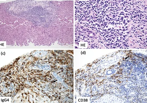
Case report: anti-GABAA receptor encephalitis after double autologous hematopoietic stem-cell transplantation for multiple myeloma
Autoimmune encephalitis associated with autoantibodies against GABAA receptors after double autologous hematopoietic stem-cell transplantation for multiple myeloma
Editor’s Choice


Featured Topics COVID-19/ Long COVID Syndrome


Support our independent journalism
If you like DiscoverMedNews, please consider supporting us and our open and independent science journalism.
Every reader contribution, regardless of the amount, is greatly appreciated.
Please read more on the Donation Page.
Thank you.
Featured Topics- Other Categories


Conferences
14TH INTERNATIONAL CONGRESS ON AUTOIMMUNITY. Ljubljana, Slovenia. 17-20 May 2024
The International Congress on Autoimmunity is the largest multidisciplinary congress that covers all aspects of related diseases, offering lectures and courses by some of the world’s most renowned experts.